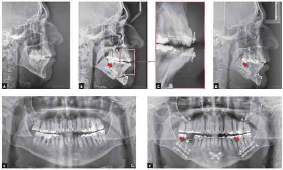
Figure 6. Cephalometric radiographs: initial (A) and immediately after surgery (B). Note that the mandibular advancement was greater than the maxillary advancement, leading to an anterior crossbite (C). This relationship was corrected by means of skeletal anchorage miniplates, which provided the necessary anchorage for lower arch retraction (arrows in B, D and F). The right lower first molar was removed before surgery (E). Panoramic radiographs: initial (E) and immediately after surgery (F).

