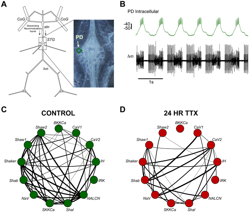
Figure 1. Network silencing disrupts ion channel mRNA relationships in single, identified PD neurons.
(A) Schematic of the Stomatogastric Nervous System from the crab, Cancer borealis. Descending modulation from the Commissural Ganglia (CoG), whose axons travel through the stomatogastric nerve (stn), is the main neuromodulatory input to the stomatogastric ganglion (STG). The pyloric dilator (PD) neuron used in this study is outlined in the photomicrograph of the STG. (B) Example of an intracellular recording of the PD neuron (top) and the corresponding extracellular recording of the lateral ventricular nerve (Ivn) that contains PD axons. Scale bar represents 10 mV. (C) Correlation network plot used to show strong relationships between 13 ion channel mRNAs in single PD neurons (n=11; series 1 control). Two nodes connected by a line represents a pairwise correlation with an r-value greater than 0.65 (the r-value where p<0.05). Line thickness increases with r-value. (D) Correlation network plot of single PD neurons silenced with tetrodotoxin for 24 hrs (n=13).

