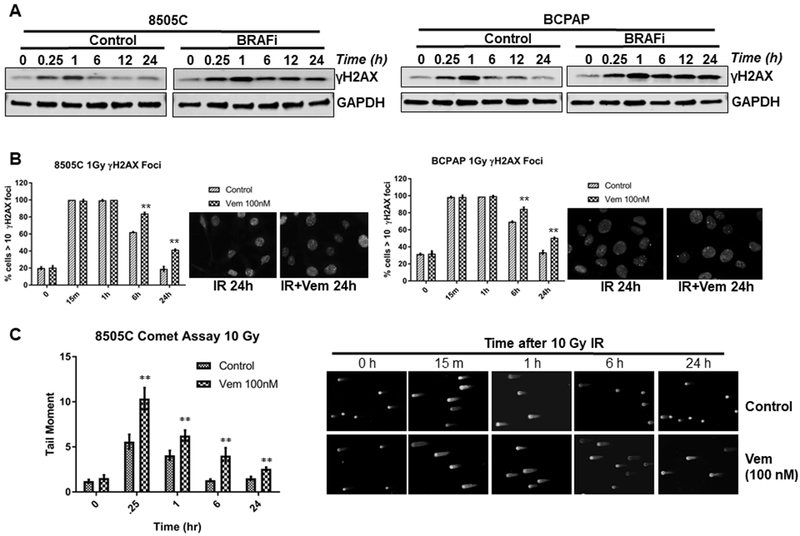
Figure 3. BRAFV600E inhibition prevents the recovery of IR-induced DNA damage in BRAFV600E mutant thyroid cancer cells.

(A) BRAFV600E mutant 8505C and BCPAP cells were cultured in media containing 100 nM vemurafenib (BRAFi) 3 hrs prior to 4Gy dose of irradiation. At the indicated time points in hours following IR, cell lysates were made for immunoblotting of γ-H2AX and GAPDH. The 0 time point indicates no IR, but treated with 100 nM vemurafenib for 3 hrs. (B) BRAFV600E mutant 8505C and BCPAP cells were cultured in media containing 100 nM vemurafenib 3 hrs prior to 1Gy dose of irradiation. At the indicated time points following IR, the cells were prepared for immunofluorescence analysis of γ-H2AX nuclear foci. Mean percentage of cells with greater than 10 γ-H2AX foci and s.e.m. at each time point after IR from two experiments of no less than 100 cells per condition are shown. Representative γ-H2AX foci of the control and vemurafenib treated at 24 h post-IR are shown. (C) DNA damage was quantified as the mean tail-moment taken from no less than 75 cells at different time points relative to 10 Gy in 8505C cells treated with/without 100 nM vemurafenib (Vem). Representative set of photomicrographs of control and vemurafenib treated comets at 24 h post-IR are shown. **p<0.01.
