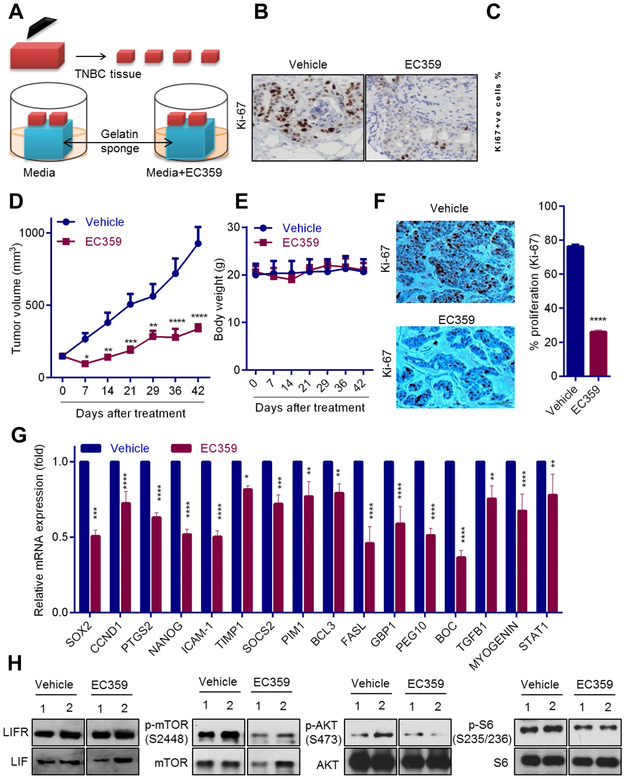
Figure 6.
EC359 decreases the growth of patient-derived explants (PDEx) ex vivo and PDX tumors in vivo. A, Schematic representation of ex vivo culture model. B, TNBC explants were treated with EC359 for 72 h and the proliferation was determined using Ki-67 immunostaining. Representative Ki-67 staining from one tumor treated with vehicle or EC359 is shown. C, The Ki67 expression in TNBC explants (n=3) is quantitated. D, TNBC PDX tumors (n=6) were treated with vehicle or EC359 (10mg/kg/s.c./3 days/week). Tumor volumes are shown in the graph. E, Body weights of vehicle and EC359 treated mice are shown. F, Ki-67 expression as a marker of proliferation was analyzed by IHC and quantitated. G, STAT3 target genes were measured by using RT-qPCR analysis (n=3). H, Status of LIFR downstream signaling was measured using western blotting (data using two different PDX tumors is shown). * P<0.05, ** P<0.01, *** P<0.001, ****p<0.0001.

