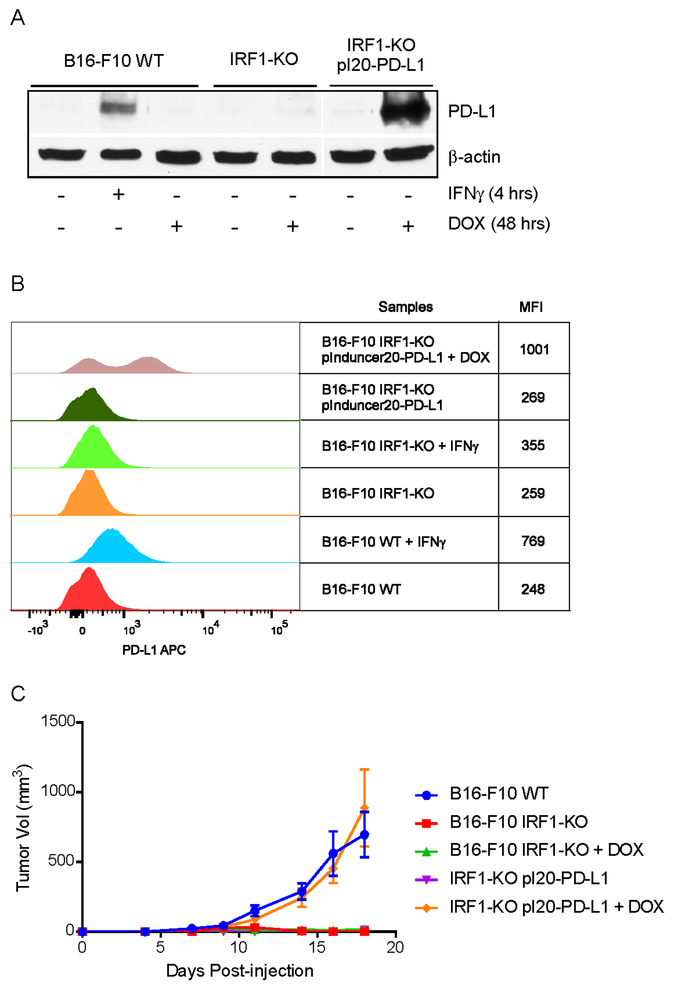
Fig. 6: DOX-induced PD-L1 expression rescues tumorigenicity of IRF1-KO cells in mice.
B16-F10 WT cells were treated with mouse IFNγ for 4 hrs for positive control. B16-F10 WT, IRF1-KO and IRF1-KO pInducer20-PD-L1 cells were treated with DOX for 48 hrs and collected for the detection of PD-L1expression. (A) Total protein expression of PD-L1 was examined by immunoblotting. (B) The cell surface expression of PD-L1 was assessed by flow cytometry. Representative histogram from twice repeated experiments are shown. (C) 5 × 10⁵ of B16-F10 WT, IRF1-KO or IRF1-KO pInducer20-PD-L1 cells were intradermally injected into C57BL/6 mice (n=5). One group of either B16-F10 IRF1-KO or IRF1-KO pInducer20-PD-L1 cell injected mice were fed with DOX containing food (200 mg/kg) from 3 days before tumor cell injection, then continued being fed with DOX containing food. Tumor growth measurements were taken with all groups of mice.

