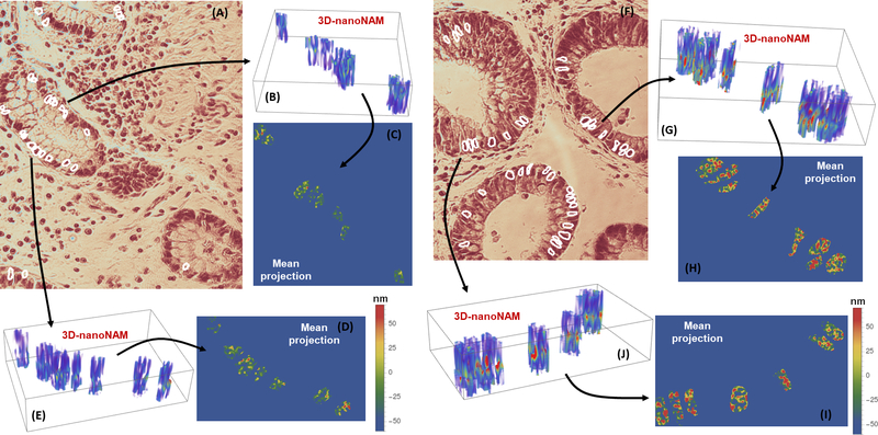
Figure 2.
nanoNAM-based visualization of 3D nuclear architecture of histologically normal appearing epithelial cell nuclei from rectal biopsies of (A-E) low-risk and (F-J) high-risk IBD colitis patients. (A and F) H&E stained tissue sample with segmented cell-nuclei of (A) low-risk and (F) high-risk patients. (B and E) nanoNAM-based 3D nuclear architecture of the unstained cell nuclei corresponding to the segmented nuclei in the stained tissue sample from low-risk patients. (G and J) depict the same for high-risk patients. In all cases, nanoNAM was performed on unstained tissue samples co-registered with the stained samples. (C and D) Mean projection of 3D nanoNAM-based nuclear architecture of cell nuclei form low-risk patients along the axial depth. (H and I) show the mean projection for high-risk patients. Although at microscopic scale the cell nuclei from both low- and high-risk patients appear histologically normal, nanoNAM-based sub-microscopic characterization shows increased alterations in mean nuclear density and associated heterogeneity in high-risk patients (redder values) compared to low-risk patients.

