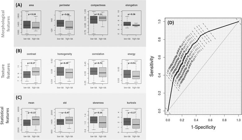
Figure 5.
Boxplots of (A) Morphological (area perimeter, compactness, elongation), (B) texture (contrast, homogeneity, correlation and energy) and (C) statistical (mean, standard deviation, skewness and kurtosis) microscopic features of histologically normal appearing cell nuclei from rectal biopsies of low-risk (dark gray) and high-risk (light gray) IBD colitis patients. (p-values have been indicated.) (D) Receiver operator characteristics (ROC) of nu-SVM based risk-classification based on the same independent validation sets used in Fig. 4, with the morphological, texture and statistical features forming the input features.

