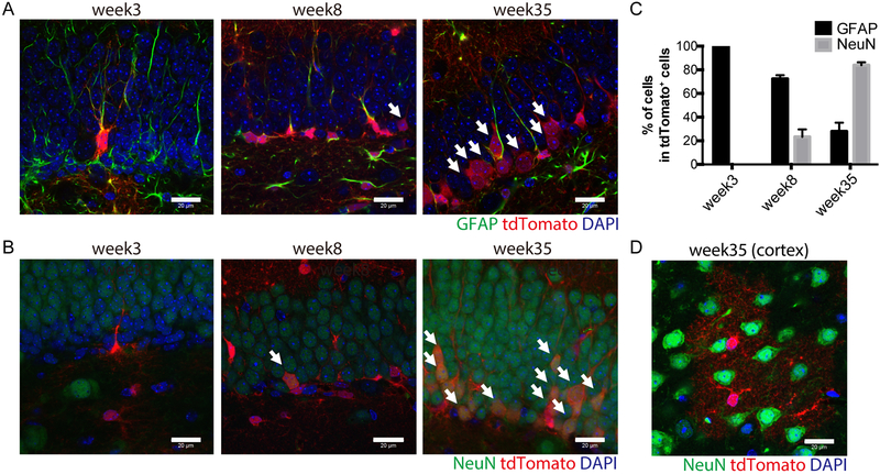
Fig. 2. FLPo-mediated recombination in GFAP-positive neural stem cells.
(A) Immunostaining analysis of GFAP (green) and tdTomato (red) expression in the subgranular zone (SGZ) of the hippocampal dendate gyrus (DG) of 3, 8 and 35-week-old GFAP-FLPo; CAG-FRT-stop-FRT-tdTomato animal. Scale bars, 20 μm. Arrows mark GFAP-negative/ tdTomato positive cells.
(B) Immunostaining analysis of NeuN (green) and tdTomato (red) expression in the SGZ of the hippocampal DG of 3, 8 and 35-week-old GFAP-FLPo; CAG-FRT-stop-FRT-tdTomato animal. Scale bars, 20 μm. Arrows mark NeuN-positive/ tdTomato positive cells.
(C) Quantified analysis of GFAP-positive and NeuN-positive cells in the SGZ of the hippocampal DG of 3, 8 and 35-week-old GFAP-FLPo; CAG-FRT-stop-FRT-tdTomato animal. Data are means with SEM. 3 animals per condition were subjected to the analysis.
(D) Immunostaining analysis of NeuN (green) and tdTomato (red) expression in the cortex of 35-week-old GFAP-FLPo; CAG-FRT-stop-FRT-tdTomato animal. Scale bars, 20 μm.

