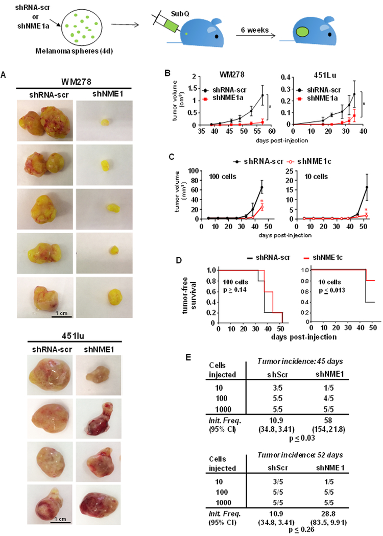
Figure 4.
NME1 promotes tumor growth properties of cells derived from melanoma sphere cultures. Melanoma sphere cells from WM278 or 451lu cell lines were cultured and transduced with lentivirus expressing either shRNA-scr or shNME-1a. Four days after viral transduction and growth in sphere culture, cells were dissociated into single cells and injected subcutaneously into the rear flank of NSG mice. A, Tumor xenografts from WM278- and 451Lu-derived cells at the experiment endpoint (5.5 wks. or tumor size > 1 cm3). B, Tumor growth curves obtained after subcutaneous xenografting of sphere cells derived from the WM278 (left) and 451Lu (right) cell lines in NSG mice. Data points represent mean ± SEM (n=5, p ≤ 0.05). Growth curves were compared using a regression ANOVA model. C, Growth of 451Lu xenografts initiated with either 100 or 10 cells. Asterisks denote means that are significantly different (p ≤ 0.05). D, Kaplan-Meier plots of tumor-free survival after subcutaneous injection of 451Lu sphere cells (100 or 10, n = 5). Differences in tumor-free survival between the treatment groups were determined using the log-rank test. E, After 52 days, mice were euthanized and tumor-initiating frequency was calculated using extreme limiting dilution analysis (ELDA), as described (28).

