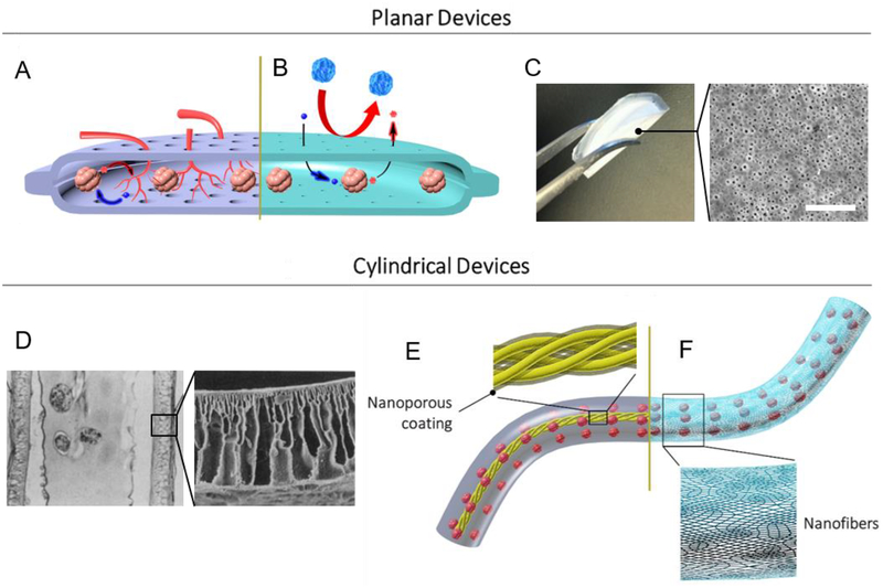
Fig. 4: Nanotechnology in Macroscopic Encapsulation and Cell Delivery Devices.
(A) Cell permissive (“open” or non-immunoisolating) macroencapsulation devices (such as PEC Direct) allow blood vessel penetration through pores from the host, supporting islet survival and regulation of glucose. Insulin (red) and glucose (blue) shown. (B) Immunoisolating macroencapsulation device with pores on the nanoscale. Host immune cells are prevented from accessing the graft, while glucose and insulin must diffuse through the permselective membrane. (C) Digital image of a folded immune-protective planar device described by Chang et al. [249] (left); top-view scanning electron microscopy image of nanoporous immunoisolating PCL membrane (right; scale bar: 200 nm; adapted with permission from [249]). (D) Microscope image of stained cross section of cylindrical hollow fiber device described by Lacy et al. [257] (left; magnification: ~41×); scanning electron microscopy image of acrylic copolymer membrane (right; magnification: ~400×; adapted with permission from [257]). (E) Thread reinforced alginate tubes use a nanoporous coating (shown in transparent gray over yellow thread) to crosslink alginate from the inside, while (F) a cylindrical nanofiber mat provides mechanical support and can function as a cell penetration resistant membrane.

