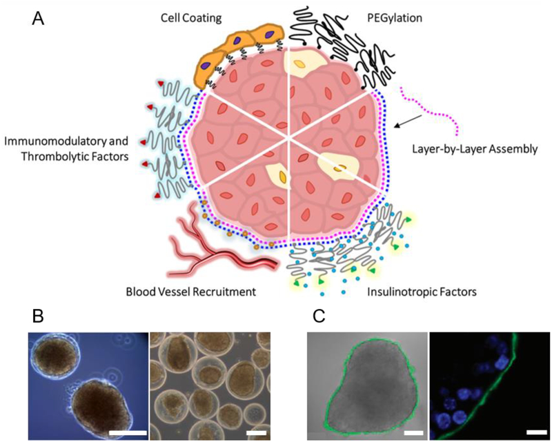
Fig. 6: Nanoencapsulation.
(A) Nano-thin coatings may be generated by (top right, proceeding clockwise): PEGylation; layer-by-layer (LBL) assembly of alternating polymer layers (e.g. polycations, pink, and polyanions, blue) deposited directly on the islet surface. Nano-coatings can be functionalized with (continuing clockwise): bioactive accessories such as insulinotropic agents including glucagon-like peptide (GLP)-1 (green; insulin shown as blue circle); blood vessel recruiting factors (e.g. heparin, orange circles); immunomodulatory and thrombolytic agents (red; e.g. soluble complement receptor (sCR)-1, thrombomodulin, urokinase, phosphorylcholine, heparin); or a cellular layer (e.g. endothelial cells, immunomodulatory cells). (B) Examples of conformal coating: phase contrast images of mouse islets conformally coated with a PEG-alginate composite gel (left) and rat islets conformally coated with a PEG-Matrigel composite gel by the method described in Manzoli et al. [339] (right; scale bars: 100 μm; adapted with permission from [339]). (C) Example of LBL nanocoating: brightfield image overlaid with confocal micrograph of 8-bilayer (PLL-g-PEG/fluorescein-labeled alginate, green) coating, fabrication described in Wilson et al. [400] (left; scale bar: 50 μm); confocal micrograph showing coating localized on peripheral islet extracellular surface (right; scale bar: 10 μm; adapted with permission from [400]).

