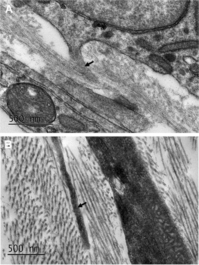
Figure 11.

Transmission electron microscopy imaging of the developing corneal stromal cells at E16 (A) and E18 (B). With maturation, the amount of collagen fibrils was seen to increase until organised orthogonal lamellae were present. There was a tendency for cell projections (black arrow) within the corneal stroma of the developing corneas at E16 and E18 to align in the same direction as some of the immediately adjacent collagen fibrils.
