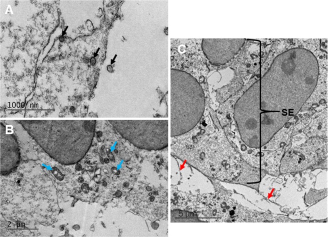
Figure 2.
Transmission electron microscopy imaging showed high resolution images of the developing cornea at E10. High resolution images of the neural crest cells that had migrated into the area of the developing cornea showed a large quantity of organelles and vesicles intracellularly and extracellularly (black arrows). (A) Synthesising organelles were also seen within the mesenchymal cells, including mitochondria, endoplasmic reticulum and Golgi apparatus (blue arrows). (B) Extracellular material (red arrows) extended from the cells of the surface ectoderm (SE) into the developing corneal stroma (C).

