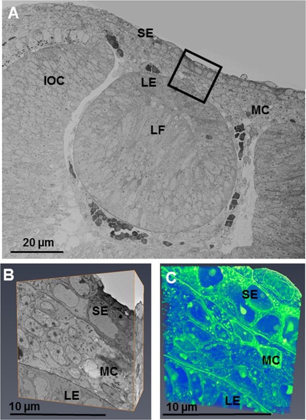
Figure 3.
The developing eye at E12 was imaged at a low magnification (×724 K) and images stitched together to show the developing lens epithelium (LE), lens fibres (LF), surface ectoderm (SE), mesenchymal cells (MC) and inner layer of the optic cup (IOC). (A) Reconstructions within the area of the black box were made following the method described in Fig. 1. (A,B) The reconstructions showed all cells in the developing corneal stroma to be in close proximity to one another, appearing to indicate a condensation of cells. (B,C) To see the cell reconstructions in more detail, please refer to Supplementary Video 2.

