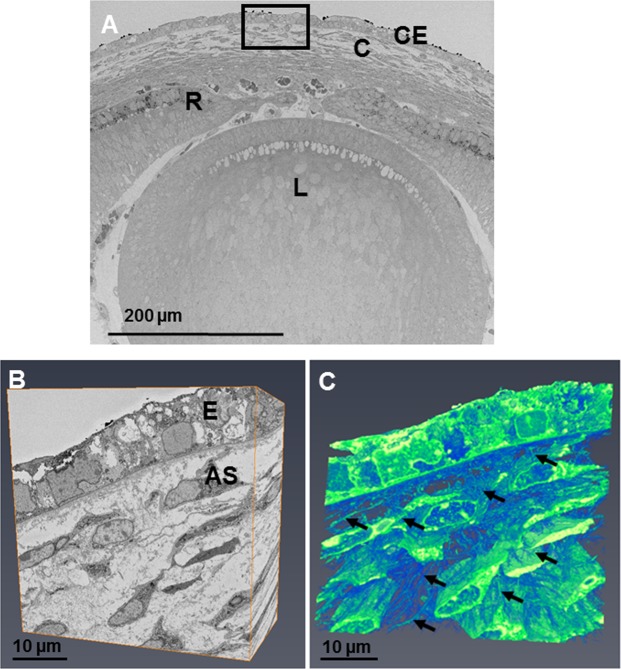
Figure 7.
The developing eye at E14 was imaged at a low magnification (×360) showing the lens (L), retina (R), cornea (C) and corneal epithelium (E). (A) Reconstructions at a high magnification within the area of the black box were made following the method described in Fig. 1. (A,B) The reconstructions showed cells within the anterior stroma that appeared stellate, surrounded by extracellular space. Reconstructions showed extensive projections from the corneal stromal cells, directed towards adjacent cells and anteriorly towards the epithelium (C) (black arrows). To see the cell reconstructions in more detail and a clearer view of the cell extensions, please refer to Supplementary Video 4.

