
Figure 8.
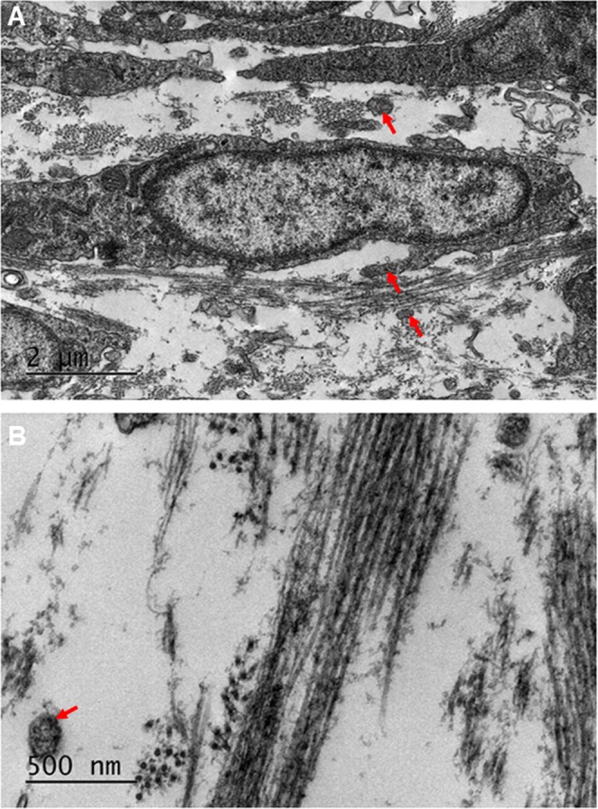
Transmission electron microscopy of the developing corneal stroma at E14 showed high resolution images of the collagen fibrils. (A,B) At lower magnification, the collagen fibrils were seen to be abundant, surrounding the cells of the corneal stroma. (A) The collagen fibrils appeared to have started to cluster within bundles, with the bundles arranged orthogonally between cells. (A) An abundance of cell projections was seen within the corneal stroma, with some associating with the collagen fibrils (A, red arrows). The collagen fibrils were imaged at a higher magnification to show the orthogonal arrangement more clearly (B).