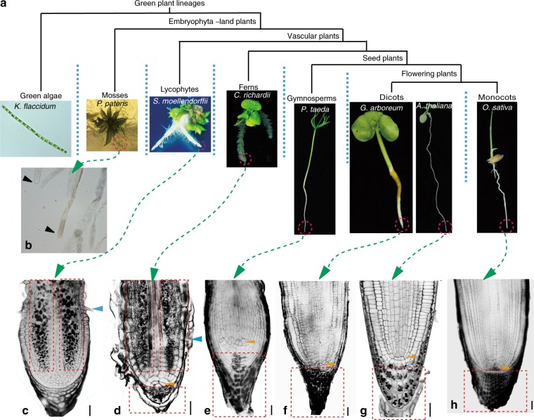
Fig. 2.
Exclusive root apex-specific amyloplast localization in seed plants. a Living representative species from different plant lineages included in the analysis (from left to right): K. flaccidum (green alga), P. patens (moss), S. moellendorffii (lycophyte), C. richardii (fern), P. taeda (gymnosperm), G. arboreum, and A. thaliana (dicots), and O. sativa (monocot). b Lugol’s staining of the rhizoid (P. patens). c–h mPS-PI staining of the root tips from S. moellendorffii (c), C. richardii (d), P. taeda (e), G. arboreum (f), A. thaliana (g), and O. sativa (h). The blue arrows indicate root hair initiation. The yellow arrows indicate the apical cell (QC-like cell) in the fern C. richardii and the QC in seed plants. The dashed red rectangles indicate the zone with amyloplasts. Scale bars, 20 µm

