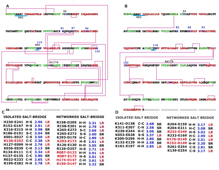
Figure 5.
Interconnection of acidic and basic partners of salt bridge on aligned sequence of hsMDH (A) and ecMDH (B) along with details of salt bridges (C for hsMDH and D for ecMDH). Buried type salt bridges are indicated by red color (C and D). Sequence color indicates secondary and coiled structures in that red indicates helix, green indicates strand and black indicates coil. SR short-ranged; LR long ranged; H helix; S strand; C coil. NAD binding regions are identified by cyan underline and NSB. Blue numbers (81, 87, 119 and 150) indicate substrate binding residues. Residues 32 and 94 indicate NAD+ binding residues. AS represent active site that takes part as proton acceptor

