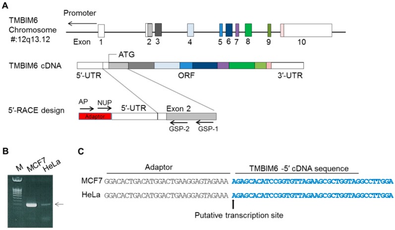
Figure 1.
Identification of the transcription start site (TSS) of the human TMBIM6 gene. (A) Schematic representation of the genome span (23.5 kb) of the TMBIM6 gene in chromosome 12q13.12. The box represents the exon positions between introns; empty box represents the UTR regions; color shaded box shows the ORF. The color box line shows the TMBIM6 cDNA with the ATG site and 10 exons. Next, 5′-RACE PCR, with adapter and nested PCR joined to the 5′ end of the cDNA and gene-specific primers GSP1 and GSP2 specific to exon 2, full length cDNA was amplified. (B) 5′-RACE PCR products were separated by gel electrophoresis. (C) Sequence analysis after the PCR product clones; the arrow shows the position of the TSS (+1) of the TMBIM6 gene.

