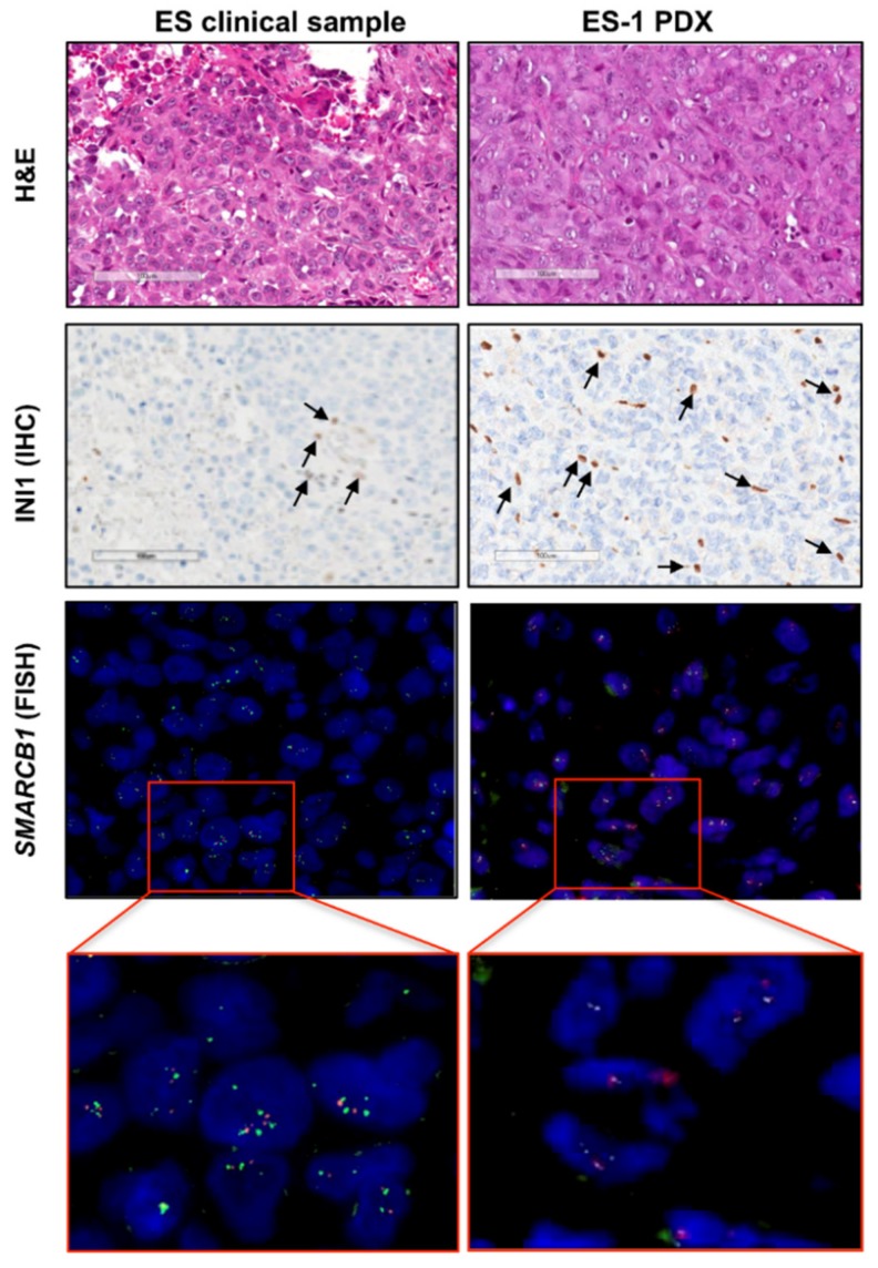
Figure 1.
Characterization of the patient-derived xenograft (PDX) model in comparison to the original clinical tumor. Representative pictures of the PDX model (epithelioid sarcoma (ES)-1 PDX) and corresponding clinical tumor (ES clinical sample) are shown. The histology was assessed on hematoxylin and eosin (H and E)-stained slides (upper panels). INI1 deficiency was detected at the protein level by INI1 immunostaining (INI1) (intermediate panels) (scale bar = 100 μm). INI1-positive intratumoral lymphocytes (internal positive control) are indicated by arrows. FISH analysis by a specific SPEC SMARCB1/22q12 Dual Color probe showed no SMARCB1 gene deletion (lower panels). The green fluorochrome-labeled probe hybridizes the human SMARCB1 gene on the chromosomal region 22q11.23; the orange fluorochrome-labeled probe hybridizes the KREMEN1 gene region in 22q12.1-q12.2. Magnification, 100×.

