Figure 2.
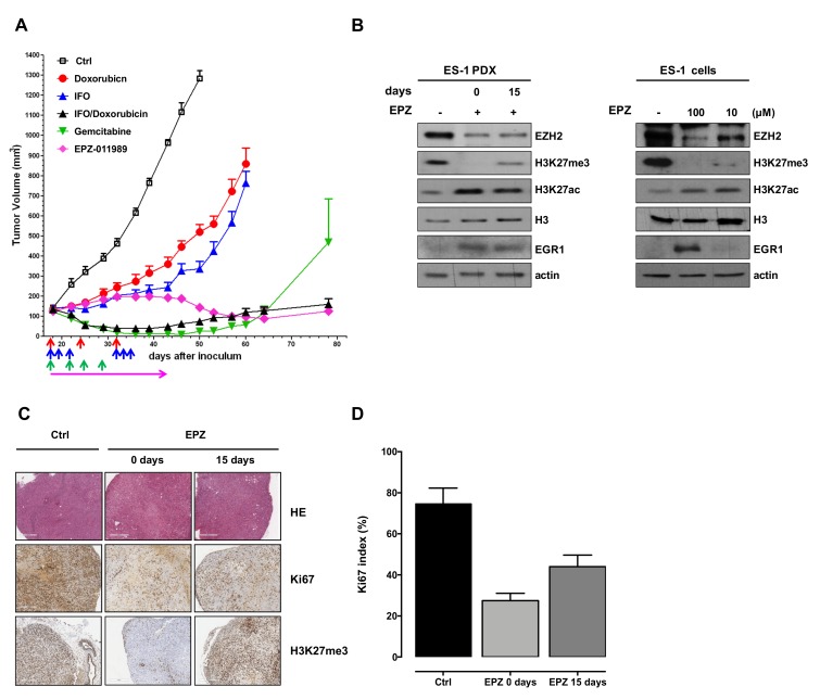
Antitumor activity and biochemical effects induced by EPZ-011989 in ES-1 PDX and derived cell line. (A) Antitumor activity of doxorubicin, ifosfamide (IFO), singly administered and in combination, gemcitabine, and EPZ-011989 (EPZ) in ES-1 PDX. Eight mice per experimental group were used. Growth curves report the average tumor volume (±SE) in control and drug-treated animal groups. The arrows in the figure indicate when the drugs were administered. (B) Western blot analysis of trimethylated and acetylated lysine 27 of histone H3 (H3K27me3 and H3K27ac, respectively), total histone H3, EZH2, and EGR1 levels in ES-1 tumors removed from untreated (−) and EPZ-011989-treated mice at different intervals from the end of treatment (left panel) and in ES-1 untreated and EPZ-011989-treated cells with different concentrations of EPZ-011989 for 96 h (right panel). Cropped images of selected proteins are shown. A representative Western blot of three independent experiments is shown. (C) Pathologic evaluation of ES-1 tumors obtained from untreated (Ctrl) and EPZ-011989-treated mice at different intervals from the end of treatment. Ki67 and H3K27me3 immunostaining of the same tumors are also presented. (scale bars = 500 µm for HE, 400 µm for Ki67 and H3K27me3). (D) Quantification of Ki67 index in untreated and EPZ-011989-treated tumors. Data are reported as mean ± SD of six different fields.

