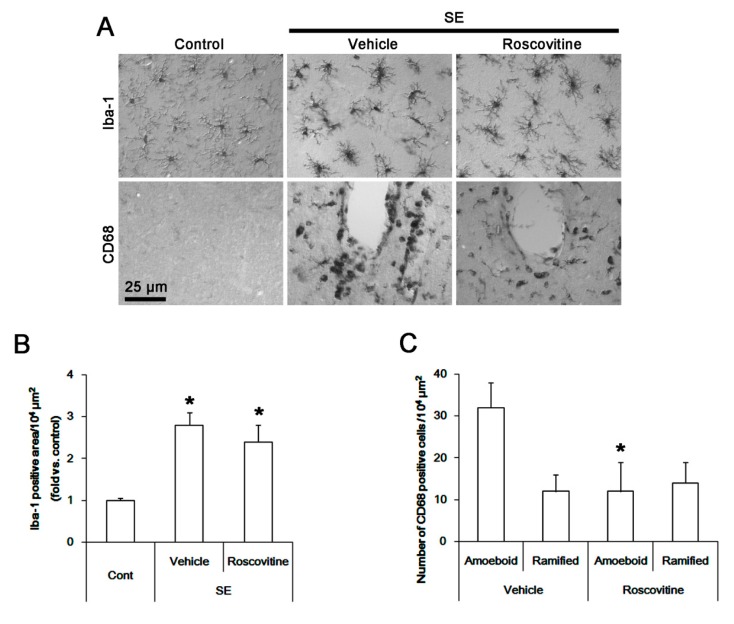
Figure 1.
The effect of roscovitine on microglia activation and monocyte infiltration in FPC following SE. Iba-1 microglia show hypertrophic morphologies with hyper-ramified processes that are covered by a lot of thorny spine following SE. In addition, CD68 cells showing amoeboid or round shapes are detected in the FPC following SE. These CD68 cells are localized in perivascular brain parenchyma. A few CD68 cells exhibited hyper-ramified shapes. Although roscovitine does not influence Iba-1 microglia transformation, it reduces the number of CD68 amoeboid cells without altering that of CD68 hyper-ramified cells. (A) Representative images for Iba-1 and CD68 positive cells. (B,C) Quantification of the effect of roscovitine on Iab-1 positive area (B) and the number of CD68 amoeboid and ramified cells (C) following SE. Error bars indicate SEM (* p < 0.05 vs. control- or vehicle-treated animals; n = 7, respectively).

