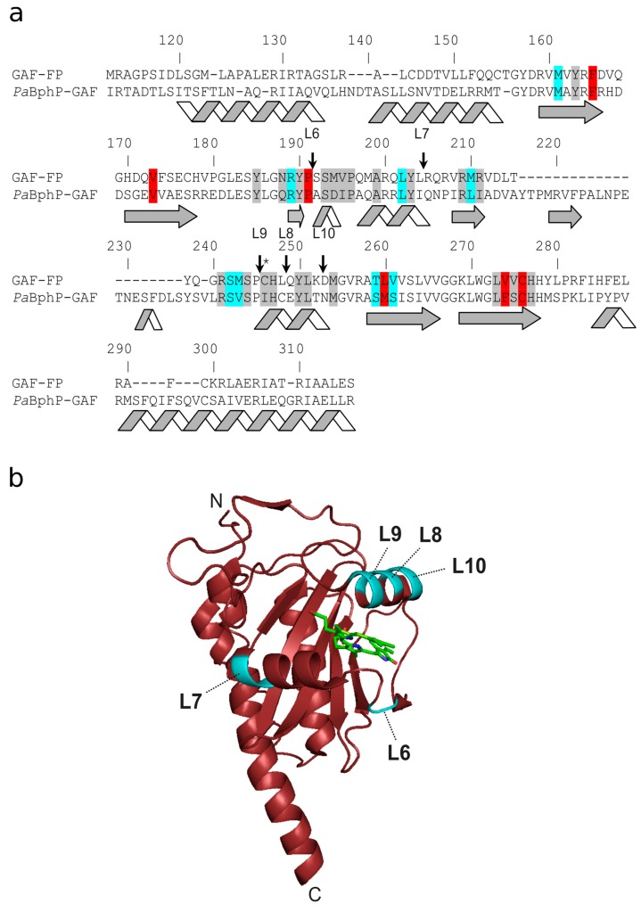
Figure 1.
Alignment of the amino acid sequences of GAF-FP and GAF domain of PaBphP (PaBphP-GAF) and representation of insertion sites of calcium binding domain. (a) Alignment numbering follows that of PaBphP. The residues which are within 4.5, 4.5−5.5, and 5.5−6.5 Å surrounding the biliverdin (BV) chromophore according to the X-ray structure of PaBphP (3C2W) are highlighted in grey, cyan, and red colors, respectively. Stars indicate Cys-residue that is covalently bound to the chromophore. Sites of insertion in the GAF-FP protein are indicated with arrows. (b) Insertion sites of calcium-binding domain are highlighted in cyan on X-ray structure of GAF domain (PDB 3C2W). The BV chromophore is shown in green.

