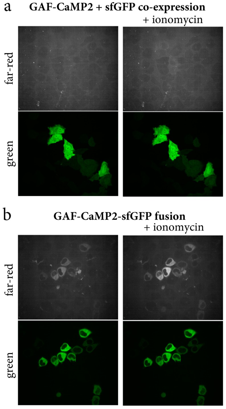
Figure 5.
Optimization of GAF-CaMP2 expression in mammalian cells. Confocal images of HeLa Kyoto cells co-expressing green sfGFP protein and far-red NES-GAF-CaMP2 indicator (a) and expressing green/far-red NES-GAF-CaMP2-sfGFP fusion (b) before and after addition of the 2.5 µM ionomycin. A total of 20 µM BV was supplied during cell’s transfection procedure 24–48 h before imaging. Green (ex. 488 nm, em. 525/50 nm) and far-red (ex. 640 nm, em. 685/40 nm) channels are shown.

