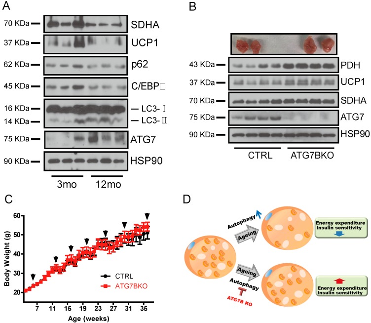
Figure 6.
Age-associated changes of BAT autophagy and mitochondrial content. (A). The relative protein levels of mitochondrial (SDHA, UCP1) and autophagy (ATG7, p62) markers in BAT of young (3-month-old) and aged (12-month-old) mice (B). Appearance of BAT depots and immunoblotting results for ATG7, UCP1 and mitochondrial-resident proteins from BAT of control and ATG7B KO mice maintained on a 60% high-fat diet (HFD). (C). Body weight chart of control (n = 7) and ATG7B KO mice (n = 9) fed 60% HFD. Arrows indicate weeks that control and ATG7B KO mice were treated with tamoxifen. (D). Schematic illustration proposing the role of brown adipocyte autophagy in age-associated decline of BAT activity.

