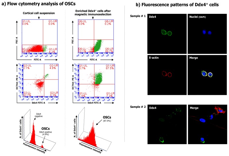
Figure 1.
Ddx4 expression on the cell population purified from the ovarian cortex.(a) Flow cytometry for Ddx4 expression measured in both total cortical cell suspension (left) and after Ddx4+ cell selection emphasized the small population extent in the cortex (4.9%) and its subsequent enrichment (87.5%). (b) Immunofluorescence for Ddx4 expression in two samples of purified Ddx4+ cells by confocal microscopy. The fluorescence patterns confirm the membrane localization of Ddx4 (FITC; green), while the cell integrity was assessed by both actin (phalloidin; red), and nuclei (DAPI; blue) staining.

