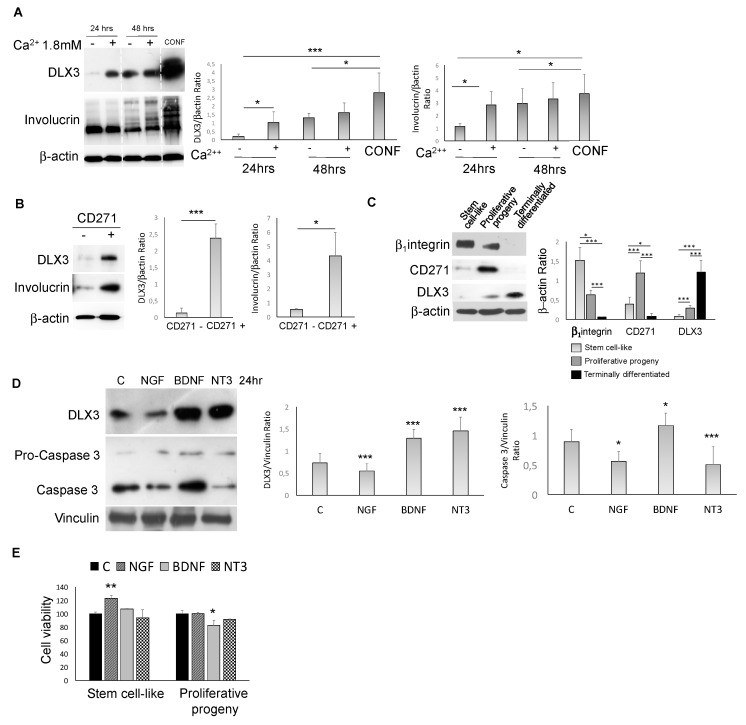
Figure 3.
CD271-DLX3 interplay in keratinocytes (A) DLX3 and involucrin expression evaluated in human keratinocytes after 24 or 48 h of 1.8 mM calcium treatment or at confluence (CONF) or (B) in CD271 negative (CD271 -) and positive (CD271 +) cells. (C) β1-integrin, CD271, and DLX3 expression in keratinocyte subpopulations isolated as described in the Materials and Methods section. (D) Expression of DLX3 and caspase 3 in human keratinocytes treated with NGF (100 ng/mL), BDNF (100 ng/mL), or NT3 (100 ng/mL), detected by Western blot. p-values have been calculated as compared to control (C in the graph) for both DLX3 and caspase 3/β–actin ratio densitometry. (E) Cell viability of stem cell-like and proliferative progeny subpopulations evalutated by MTT assay at 48 h after the addition of NGF, BDNF, or NT3 at the previously indicated concentrations. p-values have been calculated as compared to control (C in the graph). For all results, data represent the mean from three independent experiments ± SEM. For all Western blotting, densitometry was performed by ImageJ software, as described in the Materials and Methods section. * for p < 0.05, ** for p < 0.01, or *** for p < 0.001.

