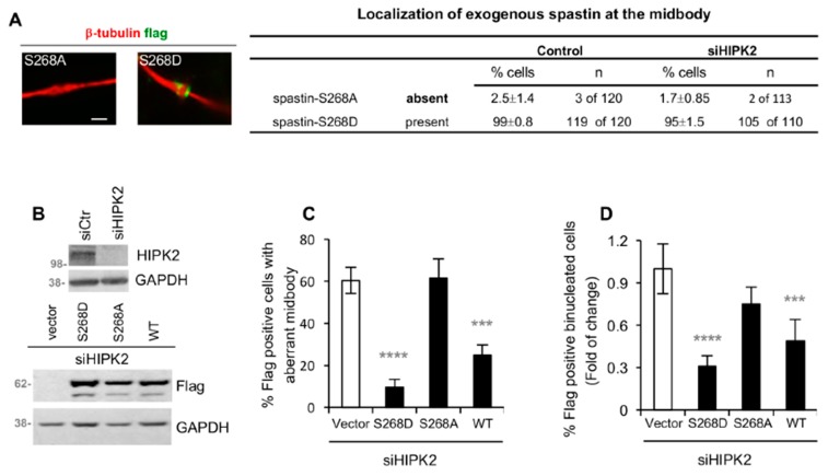
Figure 5.
Spastin-S268D restores abscission in siHIPK2 cells. (A) HeLa control cells and siHIPK2 cells were transfected with vectors expressing flag-myc-tagged spastin-S268A or -S268D and analyzed 48 h post transfection by IF after staining with DAPI, anti-β-tubulin, and anti-flag Ab to visualize midbody localization of exogenous spastin. Representative immunostainings of midbody magnification are shown in the left panels. Bar, 1 µM. Data quantification are reported, the values are mean ± SEM from three independent experiments. Similar data were obtained by using anti-myc Ab to stain exogenous spastin (data not shown). (B–D) siHIPK2 cells were transfected with flag-myc empty vector or indicated flag-myc spastin-expressing vectors 48 h post siRNA transfection performed as in Figure 1A and analyzed 48 h post vector transfection by WB and IF after staining with anti-Flag, anti-β-tubulin, and DAPI. In (B), representative WB are shown. In (C), the percentage of Flag-positive cells, showing aberrant midbodies, is reported as mean ± SEM from three independent experiments, each performed in duplicate, in which at least a total of 80 midbodies per condition were analyzed. In (D), the percentage of Flag-positive binucleated cells is reported as fold-change relative to that of vector-transfected cells in two independent experiments, in which total >2000 cells were scored. The percentages of Flag-positive binucleated cells (mean ± SEM) are the following: 4 ± 0.5, 1.95 ± 0.55, 3 ± 0.30, 1.25 ± 0.25 for siHIPK2 cells expressing empty Vector, spastin-WT, -S268D, and -S268A, respectively. ****p < 0.0001, ***p < 0.001(p = 0.0002) χ2 test.

