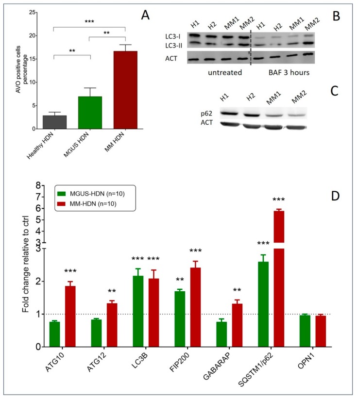
Figure 3.
Autophagy is increased in MGUS and MM high-density neutrophils (HDNs). (A) The histograms show the average numbers and standard deviations of acidic vesicular organelles (AVOs) quantified by flow cytometry using acridine orange staining, in healthy (N = 10), MGUS (N = 10), and MM HDNs (N = 10) processed within two hours from collection. ** p < 0.01, *** p < 0.001 (Student’s t-test and ANOVA test for multiple comparisons). (B) Representative immunoblot of endogenous unconjugated LC3-I to lipid-conjugated LC3-II in healthy and MM HDNs. Cells were treated for 3 h with 50 nM bafilomycin (BAF) or left untreated, lysed in 1% SDS, and analyzed by western blot with anti-LC3 Ab. ACT/actin served as a loading control throughout. (C) Representative immunoblot of p62/SQSTM1 autophagy receptor in healthy and MM HDNs. (D) Quantitative RT-PCR analysis of fold-changes of transcripts encoding autophagy machinery and responsive proteins in healthy (given 1, dashed line), MGUS, and MM-HDN. *** p < 0.0001; ** p<0.001 (Student’s t-test).

