Figure 2.
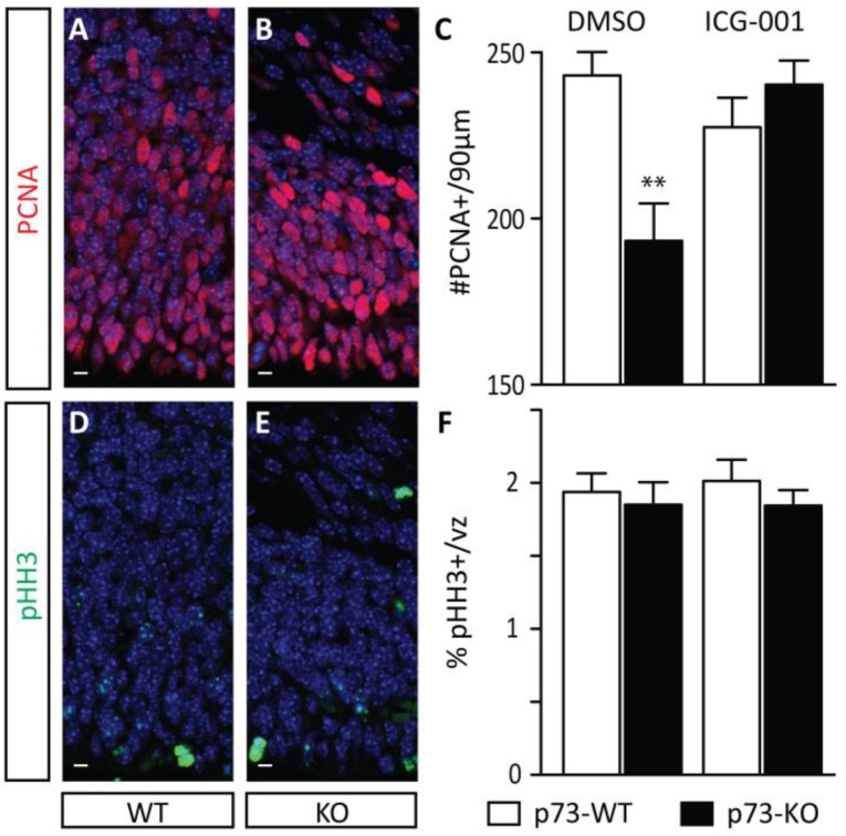
p73 knock-out mouse embryos show depletion of the germinal zone. To monitor the size of the germinal zone, PCNA immunostaining to mark proliferating cells was performed on DMSO-treated p73 wild-type (WT) (A) and knock-out (KO) (B) embryos (E17.5). A significant decrease in the number of PCNA-positive cells was observed within a 90 µm-wide column in DMSO-treated p73KO embryos compared with their WT littermates, and this decrease was rescued by treatment with CBP/β-catenin antagonist ICG-001 (C). To assess the proliferative rate of neurogenic precursors, immunostaining for phospho-histone H3-positive cells (pHH3, a marker of mitotic figures) within the PCNA-positive population was performed in DMSO-treated p73 WT (D) and KO (E) embryos (E17.5). No difference in the proliferative rate of precursors was observed as quantitated by pHH3 immunostaining (F). vz, ventricular zone. t-test. n ≥ 7. ** p < 0.01. scale bar = 10 µm.

