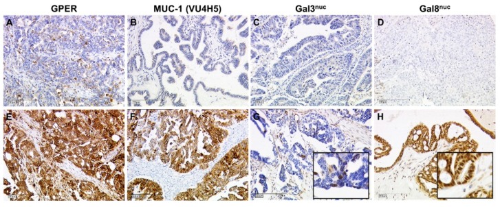
Figure 4.
Immunohistochemistry of GPER, MUC-1, and Gal-3nuc/8nuc in primary tumor tissue: GPER (A,E), MUC-1 (B,F), Gal-3nuc (C,G), and Gal-8nuc (D,H) were determined by IHC in primary OC tumor tissue. Representative images of their staining patterns scored as negative (A–D) and positive (E–H) are shown. Scale bars represent 200 µm. Inserts in G and H are magnified twice from the original image thus to illustrate nuclear staining of Gal-3 and Gal-8.

