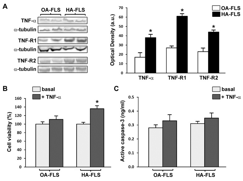
Figure 4.
Fibroblast-like synoviocytes (FLS) from patients with hemophilic arthropathy (HA-FLS) overexpress tumor necrosis factor (TNF)-α, TNF receptor 1 (TNF-R1), and TNF receptor 2 (TNF-R2) and effectively proliferate in response to TNF-α. (A) Western blotting of total protein extracts from cultured HA-FLS and osteoarthritis control FLS (OA-FLS). Representative TNF-α, TNF-R1, and TNF-R2 immunoblots are shown; α-tubulin was used as a loading control. The densitometric analysis of the bands normalized to α-tubulin is reported in the histograms. Data are mean ± SEM of optical density in arbitrary units (a.u.). * p < 0.05 vs. OA-FLS (Student’s t-test). (B) Cell viability evaluated at the basal condition or after treatment with recombinant human TNF-α (10 ng/mL) using the water-soluble tetrazolium (WST)-1 cell proliferation reagent. Cell viability in response to TNF-α is expressed as the percentage increase/decrease over the basal response for both HA-FLS and OA-FLS. Bars represent the mean ± SEM. Results are representative of three independent experiments performed with each one of the six HA-FLS and six OA-FLS lines. * p < 0.05 vs. the respective basal condition (Student’s t-test). (C) Levels of active (cleaved) caspase-3 in HA-FLS and OA-FLS, as measured by the specific enzyme-linked immunosorbent assay on cell lysates. Data are the mean ± SEM of three independent experiments performed in triplicate with each one of the six HA-FLS and six OA-FLS lines.

