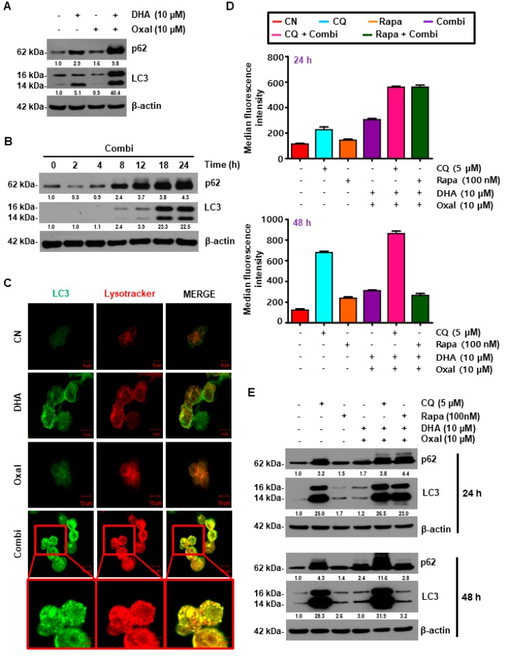
Figure 2.
DHA enhances Oxaliplatin-induced autophagy in human CRC cells. (A,B) HCT116 cells were subjected to indicated doses (A) and time (B) periods for 24 h. Then, the protein level of microtubule-associated protein 1A/1B-light chain 3 (LC3) and p62 were analyzed by immunoblotting. (C) Formation of green fluorescence protein (GFP)-LC3 puncta following exposure to Oxaliplatin and DHA was analyzed using confocal microscopy (Scale Bar, 10 μm). (D,E) HCT116 cells were treated with Oxaliplatin and DHA in the absence or presence of chloroquine (CQ) or rapamycin for 24, and 48 h. Autophagic cells (D), and autophagic markers (E) were detected using immunoblotting and a flow cytometer with an autophagy detection kit.

