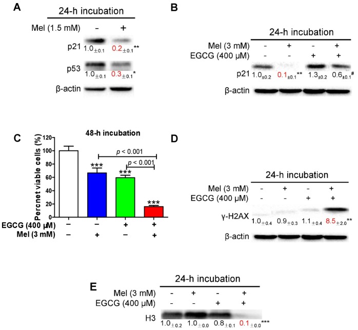
Figure 6.
Influence of melatonin, EGCG and the combination on cell viability, p21, γ-H2AX and histone H3 in HepG2 cells. (A) Influence of 1.5 mM melatonin on p21 and p53. (B) Influence of 3 mM melatonin, 400 μM EGCG and the combination on p21. (C) Influence of 3 mM melatonin, 400 μM EGCG and the combination on cell viability measured by MTT assay. (D) Influence of 3 mM melatonin, 400 μM EGCG and the combination on γ-H2AX. (E) Influence of 3 mM melatonin, 400 μM EGCG and the combination on histone H3 (H3). Data are presented as mean ± SEM (n = 6 in C or 3 in A, B, D, E). Compared to the control, * p < 0.05, ** p < 0.01 and *** p < 0.001. Compared to 400 μM EGCG, # p < 0.05.

