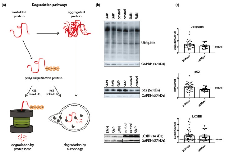
Figure 3.
Degradation pathways. (a) Misfolded proteins with polyubiquitin chains linked via lysine 48 (K48) are mainly degraded by the proteasome. Misfolded proteins carrying K63-linked polyubiquitin chains and aggregated proteins enter the autophagic pathway. Figure reproduced and adapted with permission from Dorsch, L.M. et al., Pflügers Archiv—European Journal of Physiology; published by Springer Berlin Heidelberg, 2018. (b) Representative blot images for ubiquitin, p62, and LC3BII expression. (c) Protein levels of ubiquitin and p62 did not differ between HCM and controls samples. Higher levels of LC3BII in HCMSMP (n = 38) compared to controls. There were no significant differences between HCMSMP and HCMSMN (ubiquitin, p62: n = 12; LC3BII: n = 13). Each dot in the scatter plots represents an individual sample. * p < 0.05 versus controls.

