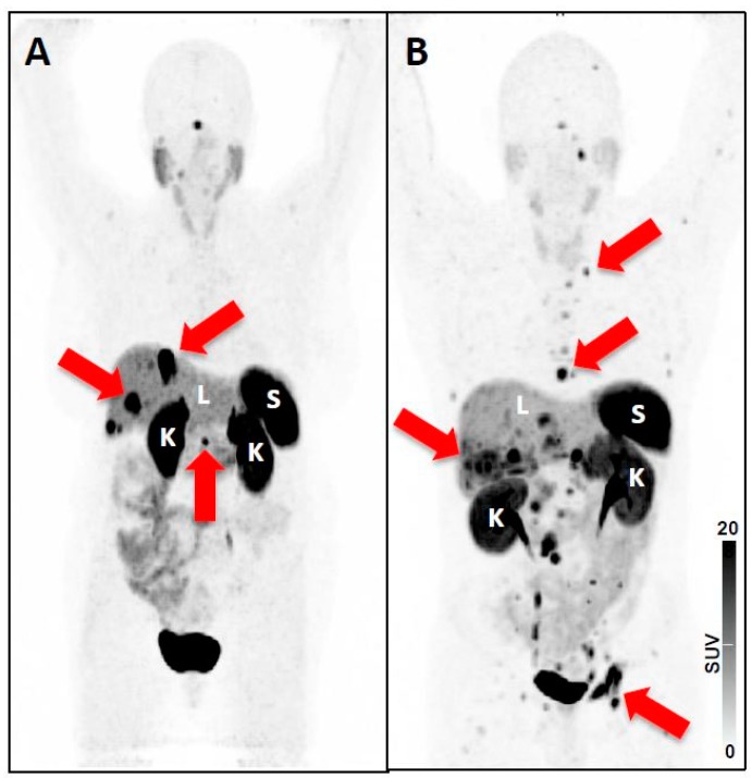
Figure 4.
68Ga-DOTATATE maximum intensity projection (MIP) of patients with (A) low tumor burden, and (B) high tumor burden. Spleen (S), liver (L) and kidneys (K) are indicated, and the threshold has been set to a SUV of 15 in both scans. Red arrows indicate tumor lesions, which can be detected on the MIP: In Patient A SSTR-positive liver lesions and the SSTR-avid tail of the pancreas are highlighted. In Patient B several thoracic lymph node metastases are shown, but also several hepatic and bone lesions (e.g., in the acetabulum) can be detected. As 68Ga-DOTATATE has been used, a moderate tumor-sink effect can be appreciated, e.g. when comparing the kidneys of both patients [42]. Nonetheless, the visual assessed uptake in normal organs (visible for liver, kidneys, and spleen) does not differ substantially among the different patients. Thus, in patients with extensive tumor involvement, subtle lesions close to normal organs or other tumor lesions with high uptake can be missed, which in turn may decrease absolute lesion detection rate.

