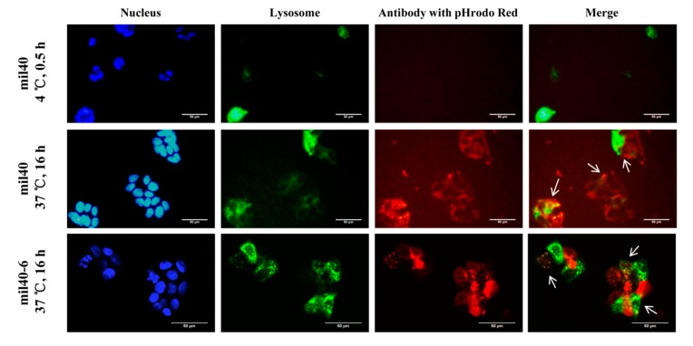
Figure 5.
Receptor-mediated internalization of ADC mil40-6 by HER2+ breast cancer cell line BT-474. Red fluorescence-labeled mil40-6 (1 μg/mL) was incubated with GFP-labeled BT-474 cells at 37 °C for 16 h while mil40-6 was endocytosed into the cell and located in lysosomes. The cell nuclei were stained with DAPI (blue), the lysosomes were labeled with a lysosomes-GFP (green), and the antobody were labeled with pHrodo Red (red). GFP: green fluorescent protein; DAPI: 4′,6-diamidino-2-phenylindole. Scale bar = 50 μm.

