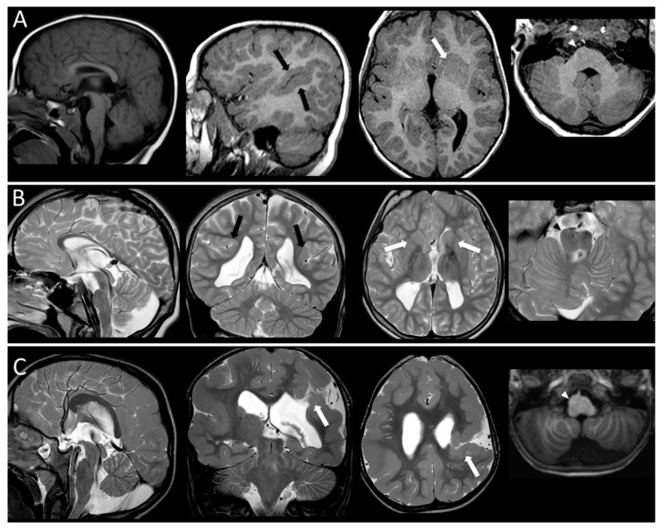
Figure 2.
MR imaging findings in three patients. Patient A [P109418] has a mutation in TUBB3 gene. T1-weighted images show a thin and short corpus callosum, perisylvian polymicrogyria (black arrow), a severe thinning/agenesis of the ALIC (white arrow) and brainstem asymmetry (arrowhead pointing at enlarged pons). Patient B [P45617] carries a mutation in TUBA1A gene. Thin corpus callosum, dysgyric cortex in perisylvian areas (black arrows), bilaterally thinned ALIC (white arrows) and pons asymmetry (arrowhead pointing at enlarged pons) are shown on T2-weighted images Patient C [P76712] carries a mutation in TUBB2B gene. He has a malformed corpus callosum and brainstem, with a thickened ponto-medullary junction and brainstem asymmetry (arrowhead pointing at enlarged medulla). He has also a complex cortical malformation with diffuse bilateral polymicrogyria and schizencephaly in the left hemisphere (arrows).

