Figure 1.
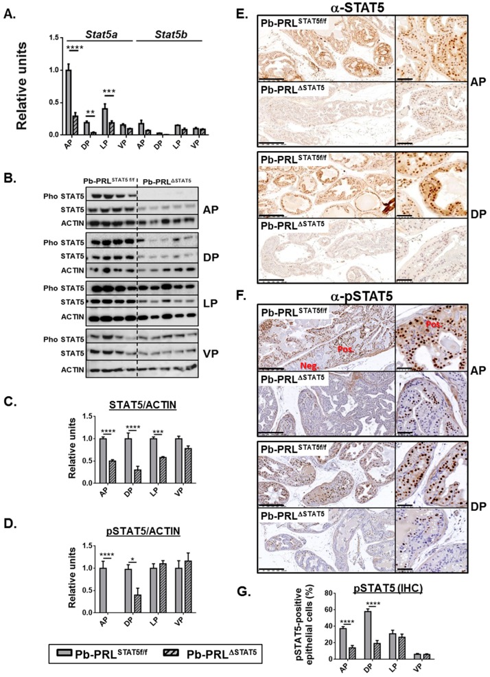
Lobe-specific pattern of STAT5 deletion in 6 month-old Pb-PRLΔSTAT5 mice. (A). Lobe-specific expression of Stat5a and Stat5b mRNA in Pb-PRLSTAT5f/f and Pb-PRLΔSTAT5 mice as determined by RT-qPCR is shown. (B–D). Lobe-specific expression and phosphorylation of STAT5 protein in Pb-PRLSTAT5f/f and Pb-PRLΔSTAT5 mice as determined by immunoblot is shown. Quantification of STAT5/ACTIN (C) and pSTAT5/ACTIN (D) was performed by densitometry and is shown as fold change versus Pb-PRLSTAT5f/f mice for each lobe. (E,F). Immunohistochemical analysis of STAT5 protein expression (E) and phosphorylation (F) in anterior (AP) and dorsal (DP) prostates of Pb-PRLSTAT5f/f and Pb-PRLΔSTAT5 mice, as indicated. In panel F, negatively (neg) next to positively (pos) immunostained areas are highlighted. See Figure S3 for the other lobes. (G). The percentage of pSTAT5-positive epithelial cells as determined using Calopix software is shown for each lobe. Statistics: Stars (* p < 0.05, ** p < 0.01, *** p < 0.001, **** p < 0.0001; idem in all figures below) denote significant differences in a repeated-measures two-way ANOVA with Sidak’s multiple comparisons. Size bars: 250 µm in large images and 50 µm in insets.

