Figure 5.
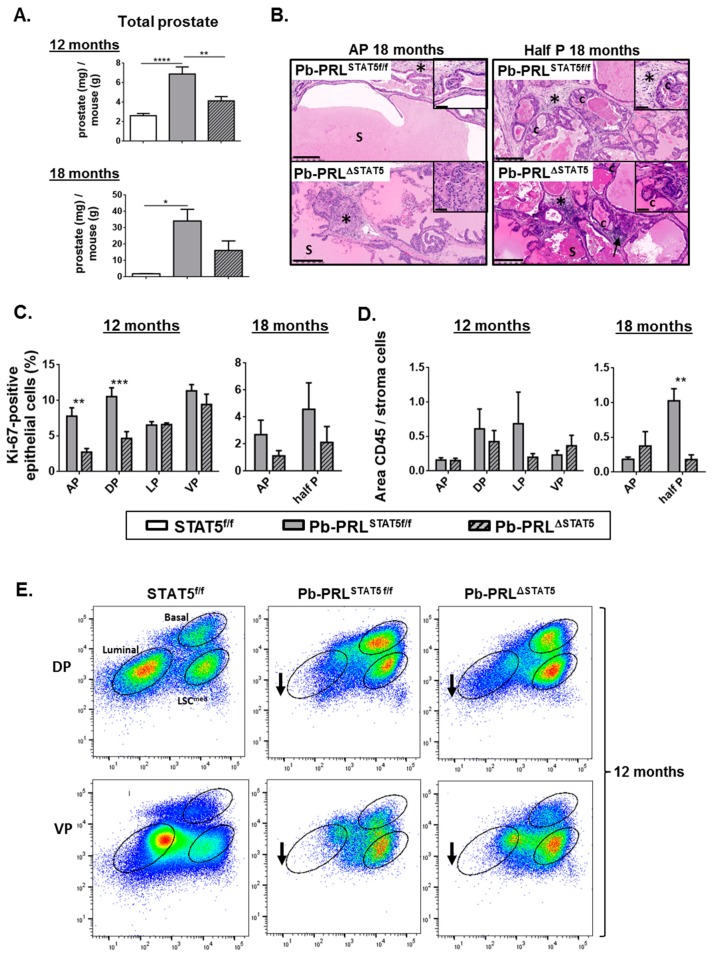
STAT5 deletion does not prevent prostate tumor progression in aged Pb-PRL mice. (A). The prostate weight in 12 and 18 month-old STAT5f/f, Pb-PRLSTAT5f/f and Pb-PRLΔSTAT5 mice is expressed as the ratio of total prostate weight normalized to the weight of corresponding animal (see Figure S5A for data per lobe). (B). Histological analysis (hematoxylin counterstaining) of prostate tumors of 18 month-old Pb-PRLSTAT5f/f and Pb-PRLΔSTAT5 mice showing similar abnormal histology including PINs, cribriform lesions (“c”), increased stromal density (stars), inflammation (arrows), and dense eosinophilic secretions (S). Sections from the anterior lobe (AP) and from fused dorsal/lateral/ventral lobes are shown. See Figure S5B for 12 month-old animals. (C). The proliferation index in prostates from 12 (all lobes) and 18 month old (anterior lobe and total prostate) mice is shown (see Figure 2 for details). (D). Inflammation was identified using CD45 immunostaining. The degree of inflammation was quantified using Calopix software and is represented as the ratio of CD45+ area versus stroma area. (E). Representative FACS profiles of dorsal (DP) and ventral (VP) prostate lobes from 12 month-old STAT5f/f, Pb-PRLSTAT5f/f and Pb-PRLΔSTAT5 mice (3–4 animals per genotype; see Figure 3A for details). Arrows show that the loss of luminal cells observed in Pb-PRLSTAT5f/f versus STAT5f/f mice was not rescued in Pb-PRLΔSTAT5 mice. Statistics: Stars denote significant differences in a repeated-measures one-way ANOVA with Tukey’s multiple comparisons (A) or two-way ANOVA with Sidak’s multiple comparisons (C). Size bars: 250 µm in large images and 50 µm in insets.

