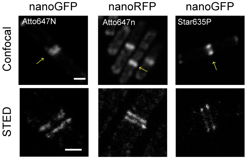
Figure 3.
Comparison of Atto647n and Star635p for bacterial STED imaging. B. subtilis cells expressing either DivIVA-GFP or DivIVA-mCherry2 were visualized using nanobodies conjugated with Atto647n or Star635p. STED images show an enlarged field of view of the object marked with an arrow in the confocal image. Scale bars 1 μm and 0.5 μm, for confocal and STED images, respectively.

