Figure 5.
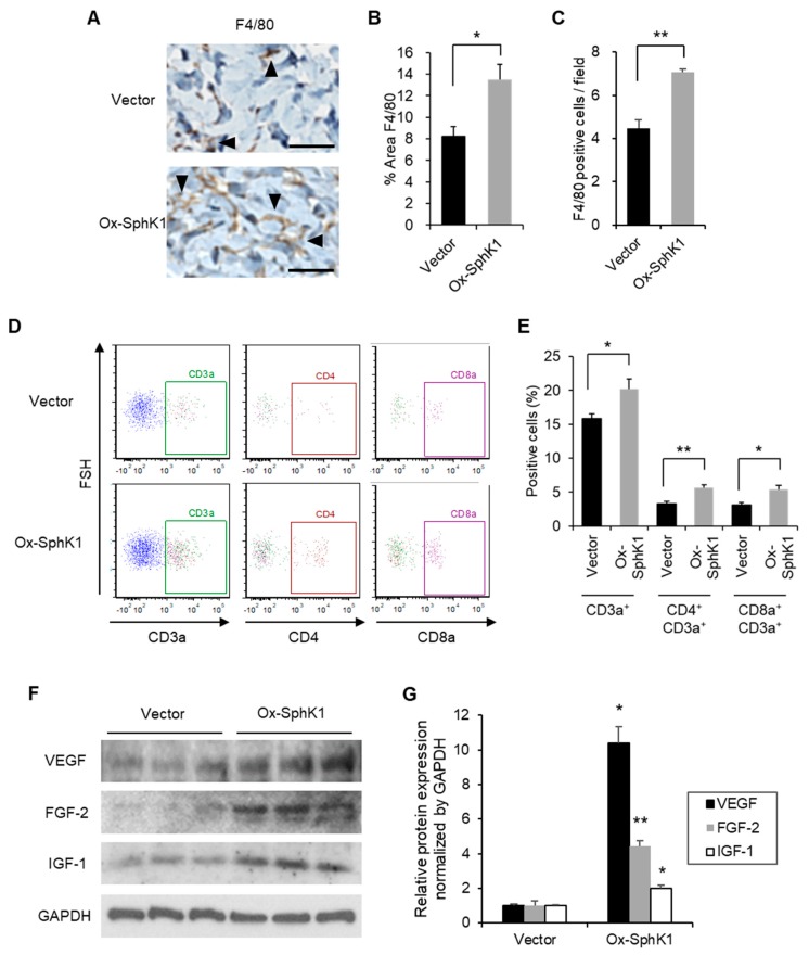
Effect of SphK1 overexpression on inflammatory cell recruitment and enhanced wound-related factors. (A) Representative images of F4/80 expression on immunohistochemistry are shown. Arrowheads indicate positive findings (scale bars: 50 µm). (B) The percentage of the wound area that is occupied by F4/80+ cells is graphed (n = 4). (C) The number of F4/80+ cells per field is graphed (n = 4). (D,E) Representative flow cytometric plots (D) and frequency of the indicated T cell populations (E) on Day 5 after wounding, as determined by flow cytometry (n = 5–8). (F) Effect of the plasmid ointment on the expression of the indicated wound healing-related factors on Day 5 after wounding, as determined by immunoblot analysis. (G) The immunoblots were quantified and the data were graphed (n = 3). All values shown in this figure represent the mean ± s.e.m. * p < 0.05, ** p < 0.01.

