Figure 1.
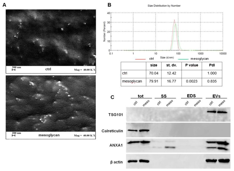
Evaluation of extracellular vesicles (EVs) isolated from keratinocytes. (A) EVs deriving from keratinocytes treated or not with mesoglycan were imaged by Field Emission-Scanning Electron Microscope (FE-SEM). Magnitude = 40,000 KX (that is 40,000,000) and scale bar = 200 nm. (B) Characterization of the EVs size distributions was found by dynamic light scattering (DLS). The table reports the EVs mean size, standard deviation, p value, referring to EVs mesoglycan distribution vs. EVs ctrl, and PdI. The experiments were performed in triplicate. (C) Western blot using antibodies against TSG101, calreticulin, and ANXA1 on protein content of total cell lysates, conditioned medium (SS), EV-depleted extracellular fractions (EDS) and EVs fractions extracted from keratinocytes treated or not with mesoglycan. Protein normalization and the check of the sample quality were performed on β-actin levels.

