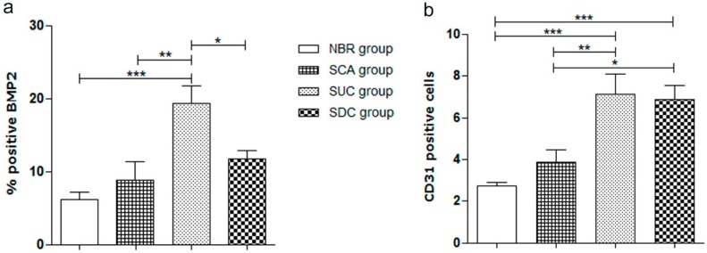
Figure 7.
Quantification of positive anti-BMP-2 (bone morphogenetic protein-2); and anti-CD31 (blood vessel) in the bone defect area after 60 days for all considered groups. NBR—animals submitted to Natural Bone Repair without a scaffold; SCA—animals submitted to PCL SCAffold without cells; SUC—animals submitted to PCL Scaffolds with Undifferentiated Cells (hADSCs cultivated only in basic media); and SDC—animals submitted to PCL scaffolds with Differentiated Cells (hADSCs cultivated in osteogenic media). The values were compared using ANOVA and Tukey’s post-test (* p < 0.05; ** p < 0.01; *** p < 0.001). Results expressed as mean ± SEM.

