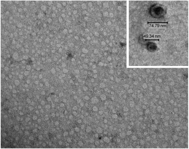
Figure 1.
Salivary EVs characterization. A representative transmission electron microscopy image of EVs isolated by a charge-based precipitation method, showing a carpet of vesicles in the nano-range. In the inset, the bars indicate the size of the extracellular vesicles (EVs). The preparation was stained with NanoVan (JEOL Jem-1010 electron microscope, original magnification ×75,000; inset ×150,000).

