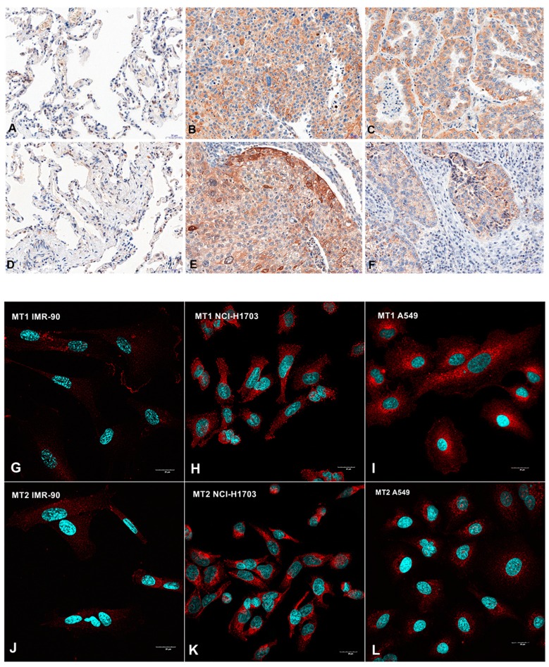
Figure 1.
Immunohistochemical (A–F) and confocal (G–L) images showing membranous/cytoplasmic expression of melatonin receptors MT1 (A–C; G–I) and MT2 (D–F; J–L) in non-malignant lung tissue (NMLT) (A,D) and human lung fibroblast (G,J), cancer cells of squamous cell carcinomas (B,E,H,K) and adenocarcinomas (C,F,I,L). Magnification ×200 (A–F) and ×600 (G–L).

