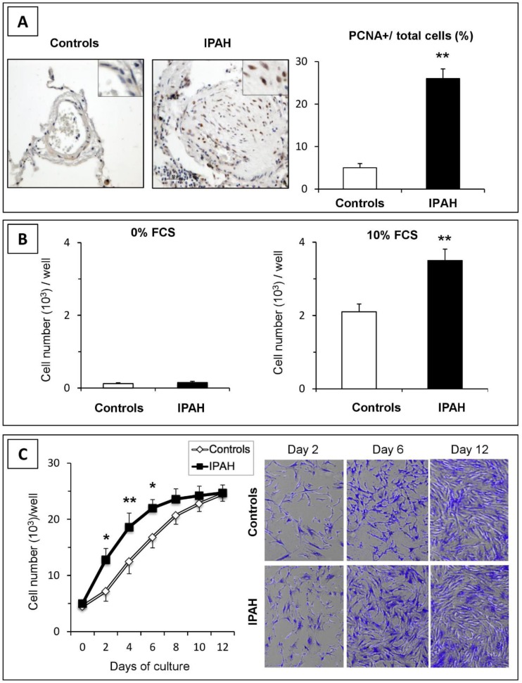
Figure 1.
Proliferation of pulmonary vascular cells in idiopathic pulmonary artery hypertension (IPAH). (A) In situ cell proliferation in pulmonary arteries from control and IPAH samples was estimated by immunohistochemistry for proliferating cell nuclear antigen (PCNA). Positive cells were more numerous in pulmonary arteries from patients with IPAH than those from controls. (B) In vitro proliferation of pulmonary artery smooth muscle cells (PASMCs) cultured without or with 10% FCS for 48 h. (C) Cellular growth curves. IPAH-PASMCs grew faster during the exponential phase of growth, but reached a similar density at confluence compared to controls (n = 6 in each group). * p < 0.05; ** p < 0.01.

