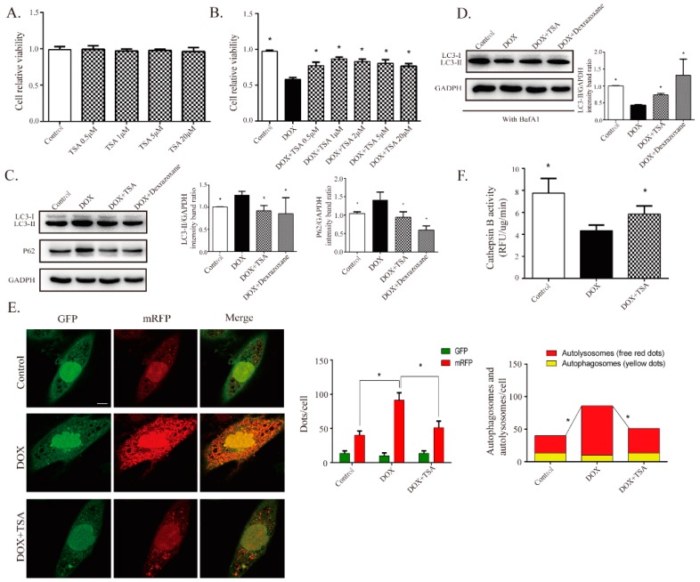
Figure 5.
TSA is shown to promote autophagosome formation and lysosomal proteolysis. (A) TSA demonstrated no cytotoxic effect on cardiomyocytes below 10 μM (n = 12). (B) TSA attenuated DOX-induced cell injury, as evidenced by increased cell viability (n = 12). Western blot results showed that TSA can reverse the expressions of LC3-II (n = 5) and P62 without (n = 5) BafA1, as seen in (C), or reverse the expression of LC3-II with (n = 5) BafA1, as seen in (D). (E) Cardiomyocytes were transfected with GFP-mRFP-LC3 to monitor changes in autophagic flux. TSA increased autophagosomes (yellow puncta) and significantly reduced autolysosomes (red puncta) (n = 30). Scale bar: 50 μm. (F) Measurement of cathepsin B activity in each group (n = 12). * p < 0.05 is significantly different as indicated, for values in the DOX group.

