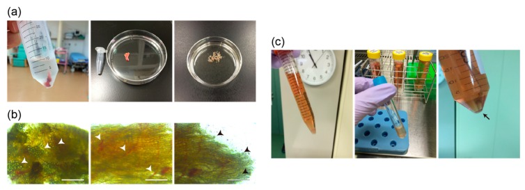
Figure 2.
The schematic representation of collecting human skeletal muscle cells obtained from extra tissues containing orbicularis oculi muscles at the time of ophthalmic surgery. (a) Surgically excised eyelid tissues soaked in cold PBS solution (left panel), an example of the actual size of the extra eyelid tissue compared with a 1.5-mL microtube (middle panel), and the obtained tissue finely chopped by scissors (right panel). (b) Morphological features of isolated tissues, mass of lipids (arrowheads in left panel), blood capillaries (arrowheads in middle panel), and disconnected skeletal muscle fibers (arrowheads in right panel). Scale bars, 200 μm. (c) Chopped samples were enzymatically treated with collagenase type 2 (left panel), then filtrated after enzymatic digestion (middle panel), and centrifuged to collect single myogenic cells (arrow, right panel).

