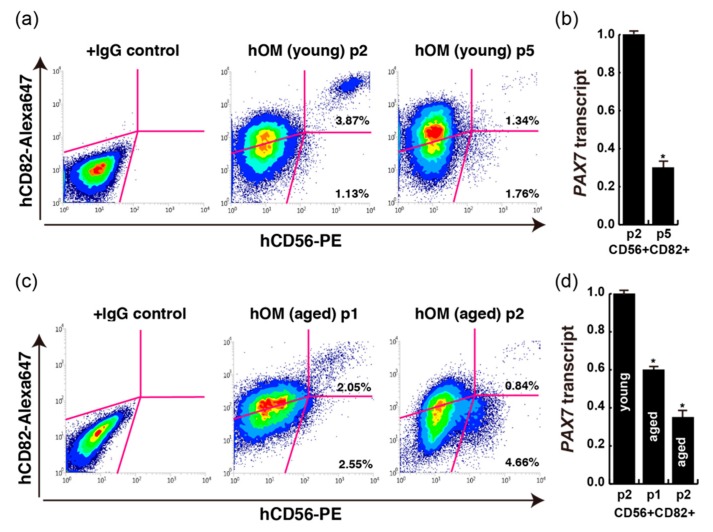
Figure 4.
FACS analyses with each primary cultured human orbicularis oculi muscle (hOM). (a) FACS data of CD56+CD82+ double positive cells from cultured double positive cells (passage 2, p2; passage 5, p5) from dissected extra eyelid tissue (14-year-old male patient; young). (b) PAX7 transcription of CD56+CD82+ sorted cells from different passage times. n = 3 independent replicates; p-values were determined by t-test from a two-tailed distribution. * p <0.01. (c) FACS data of CD56+CD82+ double positive cells from cultured double positive cells (passage 1, p1; passage 2, p2) from dissected extra eyelid tissue (74-year-old male patient; aged). (d) PAX7 transcription of CD56+CD82+ sorted cells from different age and passage samples. n = 3 independent replicates; p-values were determined by t-test from a two-tailed distribution. * p <0.01.

